
The presence of undermining can indicate shear or pulling at the wound bed and should be properly documented because the presence of preexisting damage to the subcutaneous tissues may later manifest as a visible increase in the length, width, or diameter of the wound. Undermining is another category of dead space and can be described as a hidden shelf or ledge resting just beneath the wound margin but not visible from the surface of the skin. Both tunneling and sinus formation need to be carefully assessed due to possible infection, unrelieved pressure, or the presence of foreign bodies. Sinuses may extend beyond the visible margins of the wound and are often hidden beneath the surface of the skin. Sinus formation may appear similar to tunneling but differs in that there is no exit point. The deepest area of tunneling and its location on the clock should be recorded.

Tunnels can descend deeper into the wound or to the side of the wound bed. Tunneling is generally located in the wound bed or along the wound margins and can connect one wound to another or a wound to a body cavity. In addition, the use of wound fillers to occupy dead space ensures that the wound will heal from the base to the surface without abscess formation or opportunities for deeper infection and sepsis.Ĭategories of dead space include sinus formation, tunneling, and undermining. This can be prevented by using wound fillers, such as calcium alginate or amorphous hydrogel, or in deeper wounds using gauze strips. All forms of dead space must be properly addressed during local treatment of the wound to prevent the accumulation of pathogens and exudate. Dead space represents tissue damage that is not immediately evident when inspecting visible aspects of the wound. Key characteristics that can be measured and described using the clock method are length, width, depth, and the presence of dead space. For wounds on the torso and extremities, the patient’s head is considered as the landmark for 12 o’clock and the feet are considered 6 o’clock, whereas for wounds located on the foot, the heel is considered 12 o’clock and the toes are 6 o’clock.

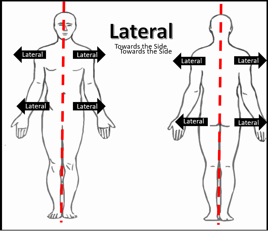
The wound is considered as a face of a clock with the position of the wound based on the standard anatomical position of the patient ( Figure 2-2). The clock method is one of the most common methods of wound measurement. The larger angle of insonation can only partly explain the differences found between LVO and RVO.Hollie Kirwan, Rose Pignataro, in Pathology and Intervention in Musculoskeletal Rehabilitation (Second Edition), 2016 Clock Method The median angle for measuring RVO is 23°.Ĭonclusion: The median angle of insonation for measuring LVO was larger than for RVO. Using vector analysis, the median angle for measuring LVO is 36° with the Doppler echo probe positioned on the thorax and 30° when positioned subcostal. The PT is directed upwards with a 53° angle (sagittal) and a 3° angle to the left (axial). The AA is directed vertically upwards to the head (sagittal) with a 40° angle to the right (coronal). Results: Forty-five patients were included with a median (range) age of 71 (2-679) days. To obtain the angle of insonation, the angle between the outflow and the assumed position of the Doppler echo probe was calculated. For each outflow the anatomical position was determined to an anatomical reference point. Methods: Magnetic resonance images of infants up till 2 years of age were explored. The aim of this study was to determine the anatomical position of the left and right ventricular outflow (Ascending Aorta, AA and pulmonary trunk, PT) and determine the angle of insonation for Doppler derived cardiac output. Differences in methodology of diameter measurements and angle of insonation could explain these findings. Background: Previous research has shown differences in Doppler derived left ventricular output (LVO) and right ventricular output (RVO), even in infants were there was no shunt.


 0 kommentar(er)
